Radiology Billing Software Built for Your Workflows
Drive efficiency and revenue with one flexible radiology platform built to handle high volumes, complex billing, and multi-site scheduling.

Tools for the Entire Radiology Workflow
Modality & PACS Integration
Our radiology software connects seamlessly with your imaging equipment and PACS, allowing you to pull results and reports into patient EHRs with ease.
MIPS & Quality Reporting
Built-in quality tracking and submission tools designed to support radiology-specific measures help you maintain compliance with healthcare regulations.
Patient Portal
Make it easy for referring providers and patients to schedule, share results, and communicate through a HIPAA-compliant patient platform.
Comprehensive & Integrated
With an all-in-one radiology solution, you’ll never have to worry about data siloes or missing functionalities. From scheduling to reporting, PracticeSuite is the only tool you need for your radiology practice.
Secure Access
Securely store patient records and communicate with them through our patient portal, earning your patients’ trust.
Superior Support
Our responsive customer support team is only ever a phone call away. PracticeSuite offers personalized assistance to ensure your practice runs smoothly using our radiology billing software.
Simpler Billing. Smoother Imaging. Faster Scheduling.

Radiology Billing Software
Handle split billing, modifiers, and imaging codes with precision. Leverage our radiology billing tools to track insurance claims, reduce denials, and get paid faster, even with high billing and claim volumes.

Radiology EHR Software
Access streamlined charting for X-rays, MRIs, CTs, ultrasounds, and more. Implement customizable templates and structured findings for more organized records.
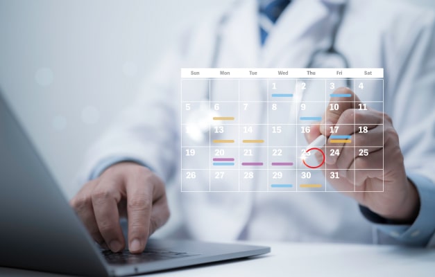
Radiology Scheduling Software
Coordinate appointments across locations, time zones, and modalities. Our radiology software also offers smart scheduling tools that help you avoid conflicts and no-shows.
PracticeSuite Reviews
Consider Us Part of Your Team
Read what medical practices, physicians, and medical billing companies have to say about the value of PracticeSuite’s medical practice management software.
“With our specialty, the appointment scheduling is very important to our organization. The PracticeSuite appointment scheduling is very customizable and gives us the ability to create our schedule based upon our needs. Our organization has found the PracticeSuite Report Central to be very easy to use. Report Central allows us to create our own reports as we need them, we like the usability. The PracticeSuite support team has been very helpful at resolving questions/issues very quickly and efficiently.”

FAQs about Radiology Billing and EHR Software
Radiology billing is complex because it involves high claim volumes, costly procedures, and strict regulatory requirements. In particular, radiology claims are often split into two parts: the technical component, which covers the use of imaging equipment and the facility, and the professional component, for the radiologist’s expert interpretation of the imaging results. Additionally, claims for high-cost imaging receive intense scrutiny from payers, making it even more difficult for radiology practices to get paid.
PracticeSuite’s radiology billing and EHR software has real-time insurance verification and prior authorization capabilities built in, so you’ll always know whether patients are covered before performing high-cost imaging.
Yes. With the help of real-time insurance verification, PracticeSuite’s radiology billing software can automatically generate accurate or good-faith cost estimates for patients and even let you know how much to charge them for their co-pay.
Yes, you can automate appointment reminders and create tailored messages for patients scheduled for specific imaging services. This means you can include the relevant prep instructions for the imaging the patient will undergo.
You take care of imaging. Our radiology software does the rest.
Made for diagnostic imaging centers, hospital-affiliated radiology teams, independent radiology practices, and more.
Schedule a Demo